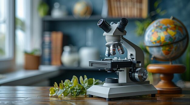
Microscopes are remarkable instruments that allow us to explore the unseen world of the very small. Whether you’re a student, researcher, or just a curious mind, understanding the components and functions of a microscope can enhance your appreciation of this essential scientific tool. This guide will walk you through the various types of microscopes, their key parts, and their applications, providing a thorough overview of how these fascinating devices work.
Overview of Microscopes
Definition and Basic Concept
A microscope is a device that magnifies small objects, making them visible to the human eye. It operates on the principle of bending light or using electron beams to enlarge images of objects that are too tiny to be seen with the naked eye.
Historical Development
Microscopy has evolved significantly since the late 16th century. Early microscopes, developed by pioneers like Zacharias Janssen and Antonie van Leeuwenhoek, laid the groundwork for modern microscopes. Advances in lens technology and the invention of electron microscopes have dramatically expanded our ability to explore the microscopic world.
Types of Microscopes
Optical Microscopes
Compound Microscopes
Compound microscopes use multiple lenses to achieve high magnification. They are commonly used in biological research to study cells, tissues, and microorganisms. These microscopes provide detailed views by focusing light through the specimen.
Stereo Microscopes
Also known as dissecting microscopes, stereo microscopes offer a three-dimensional view of specimens. They are used for examining larger objects, such as insects or plant parts, where depth perception is crucial.
Electron Microscopes
Transmission Electron Microscopes (TEM)
TEMs use a beam of electrons transmitted through a thin specimen to produce highly detailed images. They are ideal for observing internal structures of cells, viruses, and materials at an atomic scale.
Scanning Electron Microscopes (SEM)
SEMs scan the surface of a specimen with a focused beam of electrons, creating detailed three-dimensional images. They are used for studying surface structures and materials in fields like material science and nanotechnology.
Scanning Probe Microscopes
Atomic Force Microscopes (AFM)
AFMs use a sharp probe to scan the surface of a sample. They provide detailed images at the atomic level and are essential for research in nanotechnology and materials science.
Scanning Tunneling Microscopes (STM)
STMs measure the tunneling current between a sharp tip and the surface of a conductive sample. This technique allows for visualization of individual atoms and is used in advanced materials research.
Key Components of a Microscope
Base
The base is the sturdy bottom part of the microscope that supports the entire structure. It often contains the light source and provides stability during use.
Arm
The arm connects the base to the upper part of the microscope. It is crucial for handling and positioning the instrument, providing a support structure for the optical components.
Stage
The stage is where the specimen is placed for observation. It typically includes mechanical adjustments to precisely position the sample under the objective lenses.
Condenser
The condenser focuses light onto the specimen, enhancing image clarity and contrast. It plays a critical role in ensuring that the specimen is well-illuminated.
Objective Lenses
Objective lenses are mounted on a rotating nosepiece and provide various levels of magnification. Common objective lenses include 4x, 10x, 40x, and 100x, each offering different magnification powers and resolutions.
Eyepiece (Ocular Lens)
The eyepiece, or ocular lens, magnifies the image produced by the objective lenses. It is the lens you look through to view the specimen and typically has a magnification of 10x.
Illumination System
The illumination system, usually an LED or halogen lamp, provides the necessary light to view the specimen. Proper lighting is essential for clear and detailed observation.
Detailed Examination of Microscope Parts
Base
The base supports the microscope and houses the light source. It must be stable to ensure that the microscope remains steady during use. Some bases include built-in electrical components for illumination.
Arm
The arm is designed for easy handling and adjustment of the microscope. It connects the base to the optical components and should be handled carefully to avoid damaging the microscope.
Stage
The stage holds the specimen in place and can be equipped with mechanical controls for precise positioning. There are different types of stages, including plain stages and mechanical stages, each suited for various applications.
Condenser
The condenser is adjustable to control the focus and amount of light reaching the specimen. A well-adjusted condenser improves image contrast and brightness, making it easier to view details.
Objective Lenses
Objective lenses come in different magnifications and are usually mounted on a rotating nosepiece. Higher magnification lenses provide more detail but may require more precise focusing.
Eyepiece (Ocular Lens)
The eyepiece magnifies the image from the objective lenses and typically has a standard magnification of 10x. Some eyepieces come with built-in reticles for measuring specimens.
Illumination System
Modern microscopes often use LED lighting, which is energy-efficient and provides consistent illumination. The illumination system may include adjustable light intensity settings to enhance viewing conditions.
How to Properly Use a Microscope
Setting Up
Place the microscope on a stable surface and ensure that the light source is properly aligned with the condenser. Adjust the stage and prepare your specimen slide.
Adjusting the Focus
Start with the lowest magnification objective lens and use the coarse focus knob to bring the specimen into view. Once focused, switch to higher magnification lenses and use the fine focus knob for detailed adjustments.
Changing Objectives
To switch between objective lenses, rotate the nosepiece carefully. Always start with the lowest magnification lens to avoid accidental contact with the slide and ensure that the specimen is properly focused.
Cleaning and Maintenance
Regularly clean the lenses with lens paper and appropriate cleaning solutions to remove dust and smudges. Keep the microscope covered when not in use to protect it from dust and debris.
Applications of Microscopes
In Biological Research
Microscopes are essential tools in biology for studying cells, tissues, and microorganisms. They help scientists understand biological processes, disease mechanisms, and the structure of living organisms.
In Material Science
In material science, microscopes are used to examine the structure and properties of materials. They provide insights into the quality, composition, and behavior of materials at the microscopic level.
In Forensic Analysis
Forensic scientists use microscopes to analyze evidence such as hair, fibers, and particles. Microscopy can reveal crucial details that aid in criminal investigations and legal proceedings.
In Education
Microscopes are valuable educational tools that help students learn about science through hands-on experience. They allow students to explore the microscopic world and understand scientific concepts in a practical setting.
Common Issues and Troubleshooting
Blurry Images
Blurriness can occur due to misalignment of lenses or improper focusing. Ensure that the lenses are clean and correctly positioned. Adjust the focus as needed for a clear image.
Poor Illumination
If the image is dim, check the light source and adjust the condenser. Ensure that the lamp is functioning correctly and providing adequate light.
Focusing Problems
Difficulty in focusing can be caused by misaligned or dirty lenses. Check that the objective lenses are clean and properly aligned. Use the fine focus knob for precise adjustments.
Advancements in Microscope Technology
Digital Microscopy
Digital microscopes incorporate cameras and computer systems to capture and analyze images. They offer enhanced capabilities for documenting and sharing findings.
Fluorescence Microscopy
Fluorescence microscopy uses specific wavelengths of light to excite fluorescent dyes in the specimen, allowing for detailed observation of specific components within cells or materials.
Super-Resolution Microscopy
Super-resolution microscopy techniques provide images with resolution beyond the diffraction limit of light. This allows scientists to visualize details at the molecular level with unprecedented clarity.
Conclusion
Microscopes are indispensable tools that provide valuable insights into the microscopic world. Understanding their components, functions, and applications enhances our ability to explore and appreciate the tiny details of life and materials. As technology continues to advance, microscopes will remain a cornerstone of scientific discovery and education.
FAQs
What is the difference between a compound and a stereo microscope?
A compound microscope provides high magnification for observing small, thin specimens, while a stereo microscope offers a three-dimensional view of larger, thicker specimens.
How do electron microscopes differ from optical microscopes?
Electron microscopes use electron beams for imaging and offer much higher magnification and resolution compared to optical microscopes, which use light.
What are the best practices for cleaning microscope lenses?
Use lens paper and a suitable cleaning solution to gently wipe the lenses. Avoid using abrasive materials that could scratch the lenses.
How can microscopes be used in forensic science?
Microscopes are used to analyze trace evidence such as hair, fibers, and particles. They help forensic scientists identify and compare evidence to solve crimes.
What are the latest advancements in microscope technology?
Recent advancements include digital microscopy, fluorescence microscopy, and super-resolution microscopy, each offering enhanced imaging capabilities and new applications.

